Navigation auf uzh.ch
Navigation auf uzh.ch
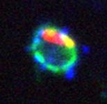
Amoeba-resistant environmental bacteria of the genus Legionella cause a potentially fatal pneumonia termed Legionnaires’ disease. The facultative intracellular bacteria grow in free-living amoebae and mammalian macrophages employing an apparently conserved mechanism. Legionella pneumophila replicates in phagocytes within a unique membrane-bound compartment, the Legionella-containing vacuole (LCV). LCVs do not fuse with bactericidal lysosomes, but interact with the endosomal, retrograde and secretory vesicle trafficking pathway, finally merging with the endoplasmic reticulum (ER). To establish the LCV, L. pneumophila employs the Icm/Dot type IV secretion system (T4SS), which translocates the amazing number of ~300 different “effector” proteins into host cells, where they subvert signal transduction and vesicle trafficking pathways.
We use macrophages and protozoa, including Acanthamoeba castellanii and the genetically tractable social soil amoeba Dictyostelium discoideum, to investigate the mechanism of LCV formation with genetic, biochemical and cell biological methods. Intact LCVs can be enriched by a simple two-step process from L. pneumophila-infected Dictyostelium amoebae producing the ER/LCV marker protein GFP-calnexin or from RAW 264.7 macrophages. The proteome of purified LCVs was determined by LC-MS/MS and revealed 670 (amoebae) or 1150 (macrophages) host proteins, respectively.
To analyze LCV formation, we currently focus on the role of host phosphoinositide (PI) lipids, small Rab GTPases and components of the retrograde trafficking pathway. Recently, we characterized L. pneumophila effectors that target PI lipids, the small GTPase Ran or the eukaryotic retromer complex.